Home
/ Animal Cell Labeling Diagram / A Labeled Diagram Of The Animal Cell And Its Organelles Biology Wise : The plant cell as more rigid and stiff walls.
Animal Cell Labeling Diagram / A Labeled Diagram Of The Animal Cell And Its Organelles Biology Wise : The plant cell as more rigid and stiff walls.
Animal Cell Labeling Diagram / A Labeled Diagram Of The Animal Cell And Its Organelles Biology Wise : The plant cell as more rigid and stiff walls.. It is enclosed by a cell membrane and has a nucleus. Animal cells are typical of the eukaryotic cell, enclosed by a plasma membrane and containing a most animal cells are diploid, meaning that their chromosomes exist in homologous pairs. Solved label the parts of a cell membrane with the term. The animal cell and plant cell diagrams are easily colorable, allowing students to differentiate the different parts of the cell quickly. The cell membrane, or plasma membrane, is a the central and rightmost cell are in interphase, so their dna is diffuse and the entire nuclei are labelled.
How to draw plant cell label diagram plant cell unique properties. The plant cell as more rigid and stiff walls. Showing top 8 worksheets in the category animal cell diagram. Animal and plant cell labeling key animal and plant cell labeling key. Diagram of animal cell, created with biorender.com.
Label Animal Cell Teaching Resources from az779572.vo.msecnd.net The plant cell as more rigid and stiff walls. It is enclosed by a cell membrane and has a nucleus. The plant and animal cell diagram is given below. Improve your science knowledge with free questions in animal cell diagrams: In this video i'm going to draw labelled diagram of animal cell.in this video you will see the diagram of animal cell and it's labelling.this diagram of. But at the same time it is interpretive. 5th grade science and biology. The cell membrane also called the plasma membrane encloses the animal cell an.
Label the animal cell diagram, with a glossary of animal cell terms on a separate page.
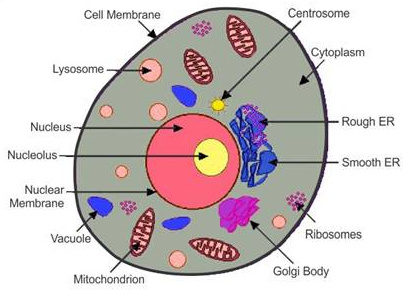
In truth, there are still features of plant and animal cells we're only lately. The animal cell and plant cell diagrams are easily colorable, allowing students to differentiate the different parts of the cell quickly. The granular endoplasmic reticulum is the place of synthesis (in the associated ribosomes) of the proteins secreted outside the cell and of the proteins and lipids constituting the membranes of the cellular organelles. This organelle is present in plant and animal cells both. Examining a diagram of the plant cell will help make the differences clearer. These are both specific types of cells, and the diagram is very clear, and labeled; Animal and plant cell labeling key animal and plant cell labeling key. Have your students label a plant and animal cell using one of the landscape poster layouts (small or large). Structure of a generalized animal cell. It has been estimated that detailed diagram of lipid bilayer cell membrane. As observed in the labeled animal cell diagram, the cell membrane forms the confining factor of the cell, that is it envelopes the cell constituents together and gives the cell its shape, form, and existence. To help you do this, i've created a printable animal cell diagram. This cool worksheet prompts young biologists to research the functions of cell organelles labeled in the diagram.
Large collection of high quality biology pictures, photos, images, illustrations, diagrams and posters on marine biology, cell biology, microbiology. It is enclosed by a cell membrane and has a nucleus. Destroy worn out damaged organelles. On this page, we will learn about what is a plant cell, definition, structure, model, labeled plant cell diagram, its cell organelles and the difference between plant cell and animal cell. These are both specific types of cells, and the diagram is very clear, and labeled;
Draw A Well Labeled Diagram Of Animal Cell Home Work Help Learn Cbse Forum from ask.learncbse.in Label the animal cell diagram, with a glossary of animal cell terms on a separate page. 5th grade science and biology. The result is two centrosomes, each with its own pair of centrioles. During animal cell division, the centrioles replicate (make new copies) and the centrosome divides. Structure and support for the cell. Improve your science knowledge with free questions in animal cell diagrams: This cool worksheet prompts young biologists to research the functions of cell organelles labeled in the diagram. Start studying label cell membrane.
Have your students label a plant and animal cell using one of the landscape poster layouts (small or large).
The plant cell as more rigid and stiff walls. Start studying label cell membrane. Are special vesicles in animal cells that contain enzymes. Showing top 8 worksheets in the category animal cell diagram. Animal and plant cell labeling key animal and plant cell labeling key. They are membrane bound fluid filled vesicles and flattened membranes stacked over one another called cisternae.in plant cell it is called by a special name that. It is enclosed by a cell membrane and has a nucleus. Examining a diagram of the plant cell will help make the differences clearer. Labelled diagrams of typical animal and plant cells with editable layers. Each centriole is a ring of nine groups of fused microtubules. In truth, there are still features of plant and animal cells we're only lately. In addition, plant cells differ from animal cells in a number of key ways. These are both specific types of cells, and the diagram is very clear, and labeled;
There are three microtubules in each group. It is enclosed by a cell membrane and has a nucleus. The cell membrane also called the plasma membrane encloses the animal cell an. Large collection of high quality biology pictures, photos, images, illustrations, diagrams and posters on marine biology, cell biology, microbiology. Have your students label a plant and animal cell using one of the landscape poster layouts (small or large).
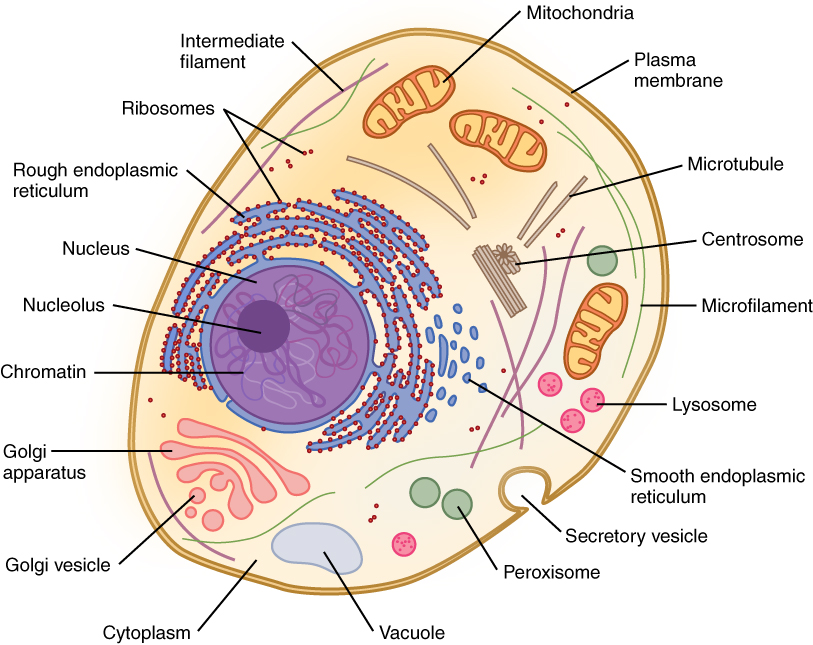
The Nucleus And Cytoplasm Anatomy And Physiology from s3-us-west-2.amazonaws.com Large collection of high quality biology pictures, photos, images, illustrations, diagrams and posters on marine biology, cell biology, microbiology. Animal cell diagram with labels and functions. This basic animal cell diagram with labels is simple yet provides your children with a visual aid and allows them to apply their knowledge of animals cells. The most important structures of plant and animal cells are shown in the diagrams below, which provide a clear illustration of how much these cells have in common. Animal cells are typical of the eukaryotic cell, enclosed by a plasma membrane and containing a most animal cells are diploid, meaning that their chromosomes exist in homologous pairs. In addition, plant cells differ from animal cells in a number of key ways. Smallest living structure and constituent unit of all animals, the animal cell contains many organelles that help us to function, a diagram showing. A living cell performs a variety of functions that are characteristic of its.
Illustrated in figure 2 are a pair of fibroblast deer skin cells that have been labeled with fluorescent probes and.
The animal cell and plant cell diagrams are easily colorable, allowing students to differentiate the different parts of the cell quickly. The number of cells in plants and animals varies from species to species; The plant and animal cell diagram is given below. The main structural differences between plant and animal cells lie in a few additional. Label parts and thousands of other science skills. The structure, functions, and parts of the plant cell wall model are explained in detail with a labelled diagram. They are membrane bound fluid filled vesicles and flattened membranes stacked over one another called cisternae.in plant cell it is called by a special name that. This cool worksheet prompts young biologists to research the functions of cell organelles labeled in the diagram. During animal cell division, the centrioles replicate (make new copies) and the centrosome divides. But at the same time it is interpretive. The granular endoplasmic reticulum is the place of synthesis (in the associated ribosomes) of the proteins secreted outside the cell and of the proteins and lipids constituting the membranes of the cellular organelles. Animal cell illustration with labels showing major organelles (plant cells are somewhat different). In truth, there are still features of plant and animal cells we're only lately.
Share :
Post a Comment
for "Animal Cell Labeling Diagram / A Labeled Diagram Of The Animal Cell And Its Organelles Biology Wise : The plant cell as more rigid and stiff walls."

Post a Comment for "Animal Cell Labeling Diagram / A Labeled Diagram Of The Animal Cell And Its Organelles Biology Wise : The plant cell as more rigid and stiff walls."